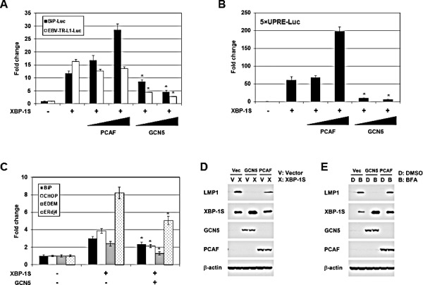
Figure 3. GCN5 negatively regulates XBP-1S-mediated transcription.

(A) HEK293 cells were transiently co-transfected with a luciferase reporter (BiP-Luc or EBV-TR-L1-Luc) and indicated expression plasmids (i.e. XBP-1S, PCAF, and GCN5). The amounts of PCAF and GCN5 plasmids were titrated at 3-fold increment. Cells transfected with an empty vector were used as a negative control. The total amounts of plasmids transfected were kept constant by adjusting the mock vector. *P<0.05 vs control (i.e. cells transfected with the XBP-1S expression plasmid). (B) HEK293 cells were transiently co-transfected with a 5×UPRE-Luc reporter and indicated expression plasmids (i.e. XBP-1S, PCAF, and GCN5). The amounts of PCAF and GCN5 plasmids were titrated at 3-fold increment. Cells transfected with an empty vector were used as a negative control. *P<0.05 vs control (i.e. cells transfected with the XBP-1S expression plasmid). (C) MCF7 cells were co-transfected with the indicated plasmid (i.e. empty, XBP-1S, or GCN5 expression vectors). The mRNAs of the XBP-1S target genes, including BiP, CHOP, EDEM, and Erdj4 were quantified by qRT-PCR. Cells transfected with an empty vector served as a negative control. *P<0.05 vs control (i.e. cells transfected with a XBP-1S expression plasmid). (D) NPC-TW01/EBV cells were co-transfected with the expression plasmids as indicated (Vec or V: an empty vector). Expression of LMP1, XBP-1S, GCN5, PCAF, and β-actin was analyzed by Western blotting 2 days post-transfection. (E) NPC-TW01/EBV cells were co-transfected with the indicated vectors. One day after transfection, the transfected cells were treated with an ER stress inducer brefeldin A (BFA, 0.1 μg/ml) for one more days, followed by Western blot analysis.
