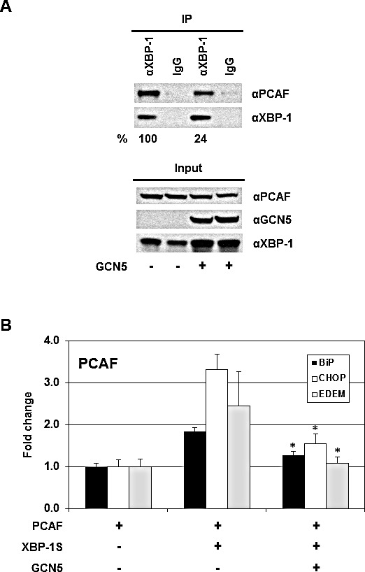
Figure 8. GCN5 competes with PCAF in binding to XBP-1S and inhibits the recruitment of PCAF to the XBP-1S target genes.

(A) 293T cells were co-transfected PCAF and XBP-1S expression vectors with or without a GCN5 plasmid. IP was performed using an anti-XBP-1S antibody, followed by immunoblotting with anti-PCAF or anti-XBP-1 antibodies. The amounts of PCAF and XBP-1S proteins immunoprecipitated by an anti-XBP-1 antibody were quantified as described under “Materials and Methods.” The XBP-1S protein precipitated in the IP against XBP-1 was used as the input to normalize the amount of PCAF protein detected in the IP. The protein inputs were also analyzed by Western blotting. (B) MCF7 cells were co-transfected with the indicated expression plasmids. ChIP was carried out followed by quantitative PCR to quantify the abundance of PCAF on the BiP, CHOP, and EDEM promoters. Cells only transfected with a PCAF vector were used as a negative control. *P<0.05 versus controls (i.e. cells co-transfected with XBP-1S and PCAF vectors).
