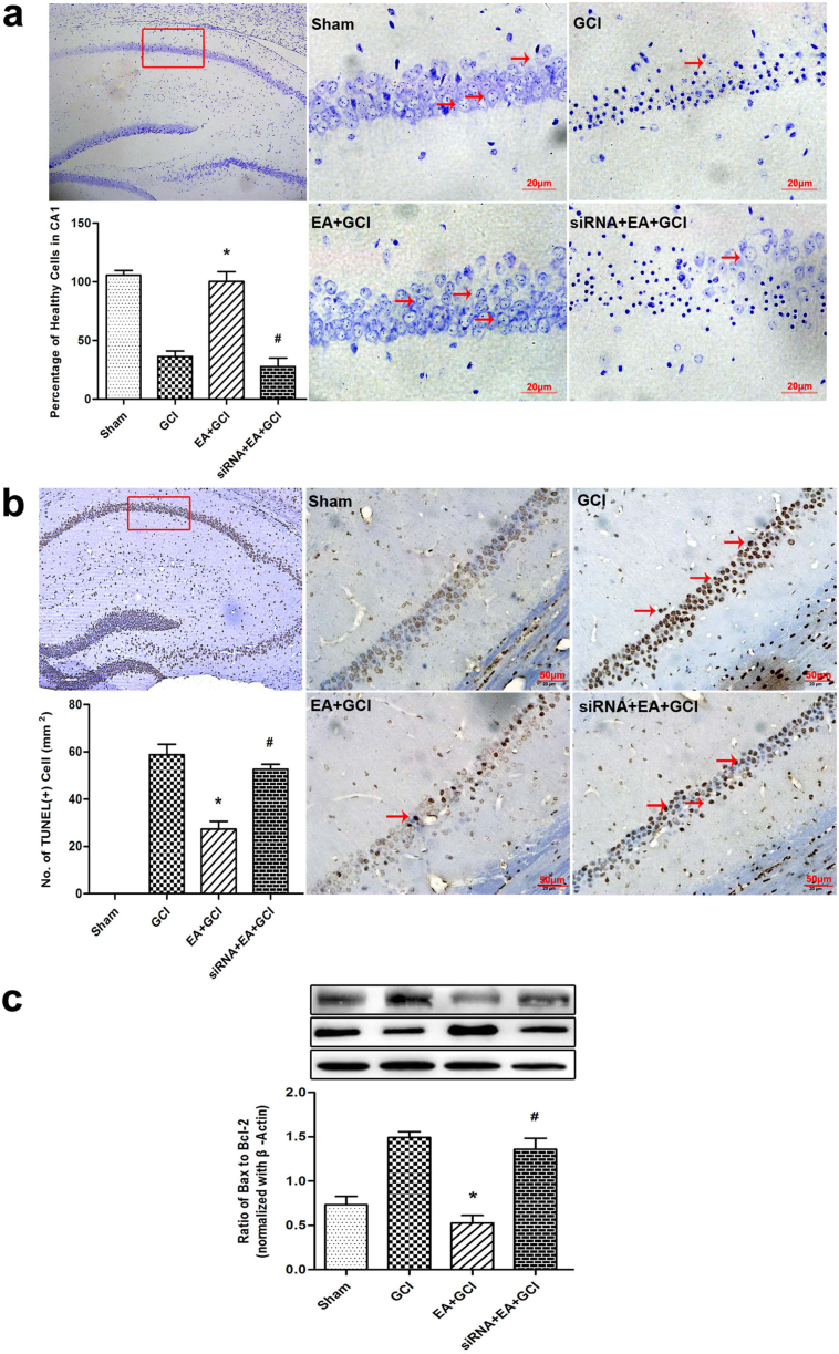
Figure 4. Neuronal survival, Neuronal apoptosis at 24 h after reperfusion in the mice with 20 min of global cerebral ischemia (n = 5).
(a)Identification of neuronal survival by Nissl-staining in rat hippocampus 24 h after reperfusion. The cell counting showed the number of viable cells in the EA+GCI group is higher compared to the GCI group (*P < 0.05 vs. GCI). GluR2 siRNA decreased the number of viable cell compared to the EA+GCI group (#P < 0.05 vs. EA+GCI). Scale bars = 20 μm. (b) Neuronal apoptosis was assessed by TUNEL staining 24 h after reperfusion in the mice. The cell counting showed an attenuation in TUNEL-positive neuronal in the hippocampus in the EA+GCI group compared to the GCI group (*P < 0.05 vs. GCI). In siRNA-treated mice, TUNEL- positive cells were significantly higher than in EA-treated mice (#P < 0.05 vs. EA+GCI). Scale bars = 50 μm. (c) The ratio of Bax/Bcl-2 expression in ischemic mice pretreated with EA or GluR2 siRNA before global cerebral ischemia was analyzed by western blot. The upper part is the photograph of Bax or Bcl-2 and its corresponding β-Actin bands. (*P < 0.05 vs. GCI; #P < 0.05 vs. EA+GCI).

