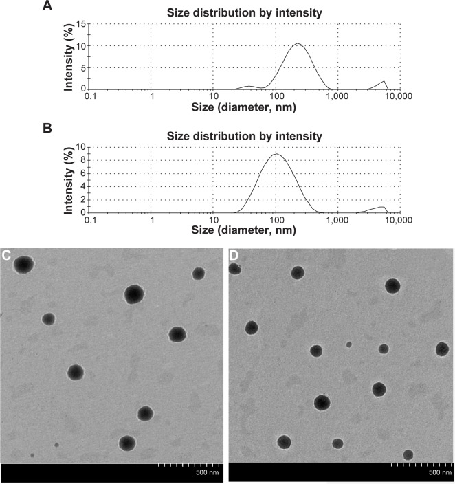
Figure 6.
Particle-size distribution and transmission electron microscopy of CIT-DOC and IT-SO-DOC.
Notes: (A) Particle-size distribution of CIT-DOC; (B) particle-size distribution of CIT-SO-DOC; (C) transmission electron microscopy of CIT-DOC; (D) transmission electron microscopy of CIT-SO-DOC. The average size of CIT-SO-DOC micelle was 100.80±7.21 nm, which was significantly lower than the 204.77±6.81 nm of the CIT-DOC group. The CIT-SO-DOC self-assembled nanomicelles seemed to be monodisperse spherical particles with smooth surfaces.
Abbreviations: CIT, circinal–icaritin; DOC, sodium deoxycholate; SO, suet oil.

