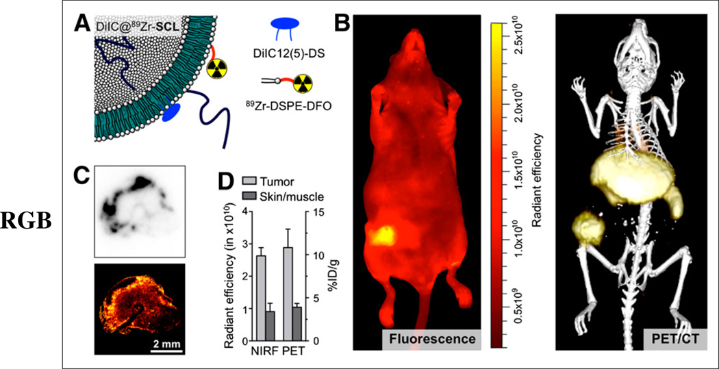
FIGURE 4.
(A) Schematic of dual-labeled liposome DiIC@89Zr-SCL. (B) Whole-body NIR fluorescence imaging (excitation wavelength = 650 nm/emission wavelength = 670 nm) (left) and 3-dimensional rendering of PET/CT fusion image (right) of same animal at 24 h after administration of DiIC@89Zr-SCL. (C) Tumor sections showing autoradiography (top) and confocal microscopy at 670 nm (bottom). (D) Comparison of NIR fluorescence (NIRF) and PET quantification measurements in tumor and skin areas (skin-to-muscle ratio for PET; n = 3).

