Figure 2.

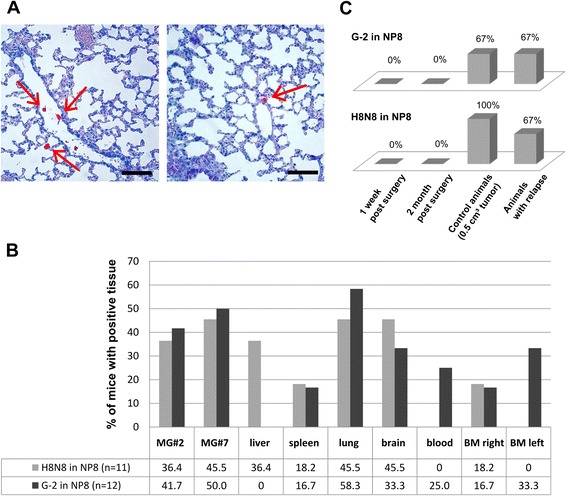
Detection of DTCs in transplanted NP8 mice. (A) Representative examples of serial lung tissue sections of mice carrying G-2 tumors at the time of sacrifice (tumor size 2 cm3), stained for T-Ag expression (red). Single positive cells (arrows) can be found in blood vessels and lung tissue. Scale bar = 200 μm. (B) Tumor cell dissemination in G-2 and H8N8 transplanted mice. NP8 mice were orthotopically transplanted with 103 H8N8 cells (n = 11) or with G-2 cells (n = 12). Different mouse tissues, blood and bone marrow (BM) were analyzed by PCR for the occurrence of DTC (HA-signal) at the time of sacrifice (tumor size of 2 cm3). Plotted is the percentage of mice with positive signals in the respective tissue, blood or bone marrow. (C) Tumor cell dissemination in G-2 and H8N8 cell transplanted mice after tumor resection. NP8 mice were orthotopically transplanted with either 103 H8N8 or G-2 cells. Tumor growth was monitored by caliper measuring. At 0.5 cm3 tumors were surgically removed and were sacrificed at 2 months (G-2: n = 5, H8N8: n = 5) and 1 week post surgery (G-2: n = 5, H8N8: n = 4). Animals with relapse (G-2: n = 6, H8N8: n = 3) and control mice (G-2: n = 3, H8N8: n = 6) were sacrificed at 0.5 cm3 tumor size. Different mouse tissues (mammary gland #7, liver, spleen, lung, brain), blood and bone marrow were analyzed by PCR for the occurrence of DTC (HA-signal). Plotted is the percentage of mice with positive signals in any of the analyzed tissues. Mice suffering a relapse of tumor growth are plotted separately.
