Figure 2.

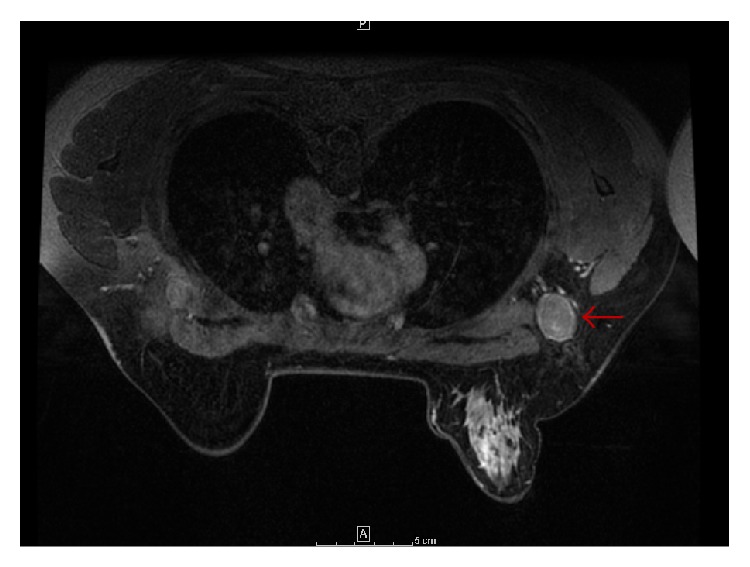
MRI breast: axial T1 image with contrast demonstrating normal breast tissue with right axillary lymphadenopathy (red arrow).

MRI breast: axial T1 image with contrast demonstrating normal breast tissue with right axillary lymphadenopathy (red arrow).