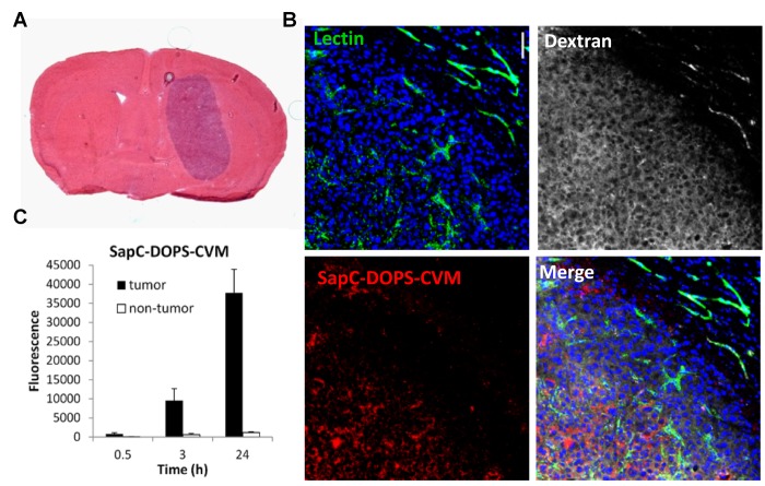
Figure 4. Intratumoral accumulation of SapC-DOPS-CVM.
A) Hematoxylin and eosin staining of a mouse brain section harboring a U87ßEGFR-Luc tumor. B) Confocal images of a GBM region and adjacent normal brain parenchyma shows specific intratumor accumulation of SapC-DOPS-CVM, 24 h after iv injection. Lectin-FITC and dextran-TRITC (MW 70 kDa) were injected before sacrifice to stain the vasculature and assess vascular permeability, respectively. C) Quantification of SapC-DOPS-CVM fluorescence from images like those shown in B.

