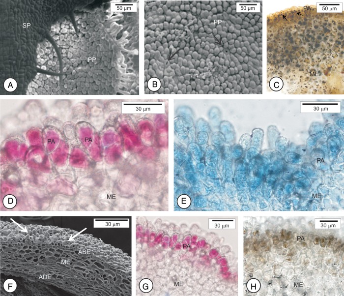
Figure 3.
Scanning electron micrographs (A, B, F) and photomicrographs (C–E, G, H) of osmophores in different developmental stages of flower buds of sweet orange (C. sinensis ‘Valência’). (A–C) Flower buds 3 mm long. The region exposed is composed of papillary cells among which the stomata occur (arrow in B). (C) Note starch accumulation in the papillary cells. (D and E) Buds 8 mm long. Papillary cells stained with neutral red dye (D) and with Nile blue sulfate (E). (F–H) buds 12 mm long. (F) Papillary cells (arrows) that react positively to neutral red dye (G) and ferric chloride (H). ABE, abaxial epidermis; ADE, adaxial epidermis; ME, mesophyll; PA, papillae; PP, petal primordia; SP, sepal primordia.

