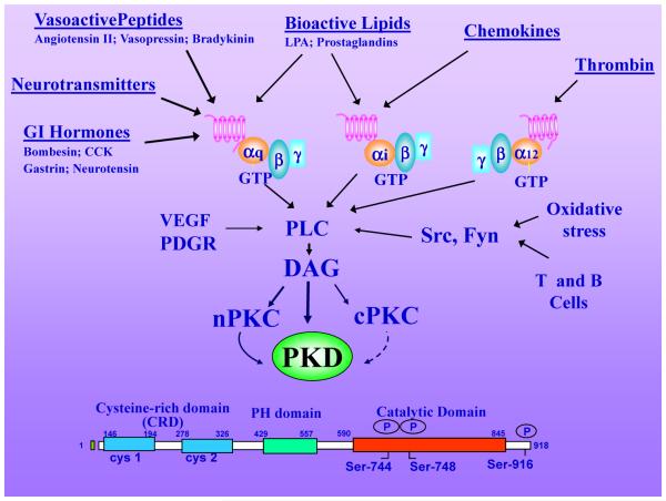
Figure 1. PKDs activation by multiple stimuli.
Hormones, growth factors, neurotransmitters, chemokines, bioactive lipids, proteases and oxidative stress induce PLC-mediated hydrolysis of phosphatidylinositol 4,5-biphosphate (PIP2) to produce DAG at the plasma membrane, which in turn mediates the translocation of inactive PKDs from the cytosol to that cellular compartment. DAG also recruits, and simultaneously activates, novel PKCs to the plasma membrane which mediate transphosphorylation of PKD1 on Ser744 (in mouse PKD1). DAG and PKC-mediated transphosphorylation of PKD act synergistically to promote PKD catalytic activation and autophosphorylation on Ser748. The modular structure of PKD (mouse PKD1) is illustrated as an example of the PKD family. PKD1 is the most studied member of the family and its knockout induces embryonic lethality. Further details are provided in the text.

