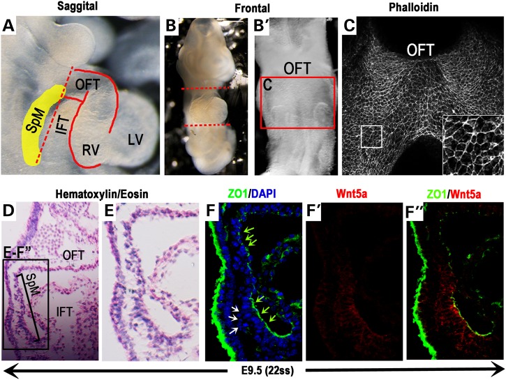
Figure 1.
SHF cells in the SpM gain epithelial character and display caudally restricted Wnt5a expression. To analyze the cellular character of SHF cells located in the SpM (yellow shaded area in A), hearts of E9.5 mouse embryos (∼22 somite stage) were removed along the dotted lines (A & B). The SpM region behind the heart tube (red box in B′) was stained with phalloidin and imaged ventrally (C). Inset in C shows magnified view of the boxed area. (D) H&E stained sagittal section of E9.5 embryo and enlarged view (black box in D) shown in (E). Immunostaining of adjacent sections shows that SHF progenitors in this region are organized as an epithelial-like sheet with tight junction marker ZO1 localized at their apical cell–cell junctions (F, green arrows). At the very caudal end of the SpM nearing the IFT, however, ZO1 expression is missing in groups of loosely packed, multi-layered SHF cells behind this epithelial sheet (white arrows in F). (F′ & F″) Co-immunostaining with an anti-Wnt5a antibody shows that Wnt5a protein is distributed in a highly restricted fashion in the caudal SpM. IFT, inflow tract; OFT, outflow tract; SpM, splanchnic mesoderm.

