Figure 4.
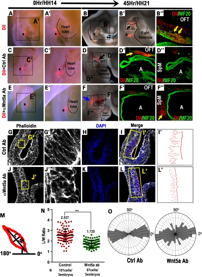
Chick SHF progenitors in the caudal SpM are deployed to the inferior OFT in a Wnt5a-dependent fashion. DiI was injected into the caudal SpM of HH14 chick embryos (asterisk in A and A′). After culturing for 45 h to HH21, DiI-labeled cells were observed in the inferior OFT region (B and B′). Sagittal sections confirmed that DiI-labeled cells (red) were present in the inferior OFT myocardial wall and co-expressed myocardial marker MF20 (green, yellow arrows in B″). Asterisks in B and B′ denote the injection site that occasionally retains some DiI labeling. (C–F″) HH14 chick embryos were co-injected with a mixture of control rat IgG and DiI (asterisk in C and C′), or anti-Wnt5a IgG and DiI (asterisk in E and E′), and harvested and analyzed at HH21. In control-injected embryos, DiI-labeled cells (red) were observed in the inferior OFT myocardium (D and D′), but were absent from the SpM region (D″). In anti-Wnt5a IgG-injected embryos, however, a large number of DiI-labeled cells were retained in SpM region behind the heart (blue arrow in F, yellow arrow in F″), and only few DiI-labeled cells were present in the OFT (F′). (G–O) HH14 embryos in which control or anti-Wnt5a IgG were injected into the caudal SpM were harvested 15 h later to assess cellular morphology in the SpM. Sagittal sectioning and phalloidin staining showed that in anti-Wnt5a IgG-injected embryos, SHF cells in the caudal SpM displayed diminished and disorganized actin polymerization (compare G′ and J′). In control-injected embryos, SHF cells in the SpM are largely elongated perpendicular to the plane of the SpM and along the dorsal-ventral (D–V) axis (G and G′); whereas in anti-Wnt5a IgG-injected embryos, SHF cells in the SpM appeared to be more rounded and randomly oriented (J and J′). To quantify cell polarity, lines were drawn along the long axis of each cell (I and L) and represented in (I′) and (L′). Length to width ratios (LWR) and the angularity of each SHF cell in the SpM were calculated as depicted (M). (N) Measurement of the LWR in SHF cells in control or Wnt5a-injected embryos. (O) Rose diagrams depicted that control SHF cells were largely aligned perpendicular to the plane of the SpM (90°) and along the D–V axis of the embryo (0°), whereas in anti-Wnt5a IgG-injected embryos, SHF cells displayed randomized orientation. A, atrium; HH, Hamburger–Hamilton stages; LWR, length-to-width ratio; OFT, outflow tract; SpM, splanchnic mesoderm.

