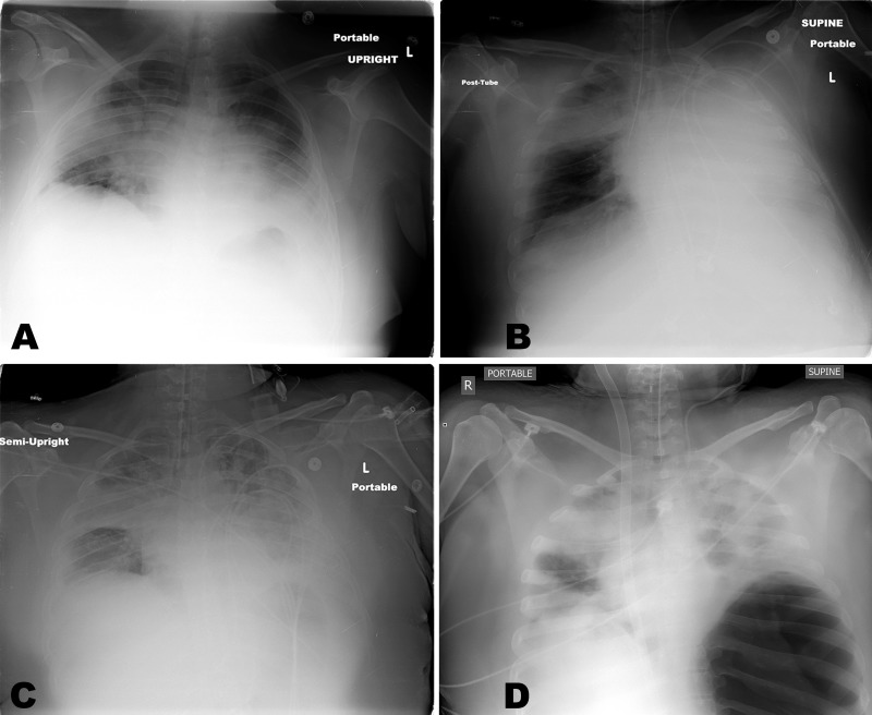
Figure 1.
Series of the chest x-rays of the patient. Initial radiograph upon presentation showed bilateral infiltrates (1A). Post intubation x-ray shows endotracheal tube in the right main stem bronchus (1B). After repositioning the endotracheal tube, there is the progression of the bilateral infiltrations (1C). Post ECMO cannulation at arrival to our facility, there is a large loculated pneumothorax in the left thorax (1D).
ECMO = extracorporeal membrane oxygenation.

