. 2014 Nov 26;3:e03579. doi: 10.7554/eLife.03579

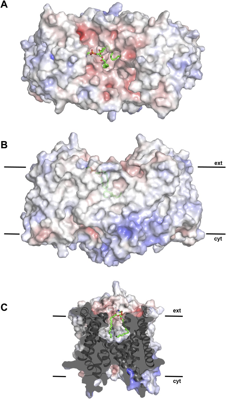
Figure 2. Hydrophobic extracellular cavity with bound lipid.

(A) One lipid molecule (PE, green) in the cavity between the two protomers in the dimer contributes to the hydrophobic contacts across the dimer interface. The extracellular surface is slightly negatively charged. (B) The alkyl chain of the lipid extends to the center of the molecule. (C) The lipid-facing surface of the central cavity is mainly hydrophobic. The surface potential was calculated at pH 7.0 by APBS.

DOI: http://dx.doi.org/10.7554/eLife.03579.010

Figure 2.

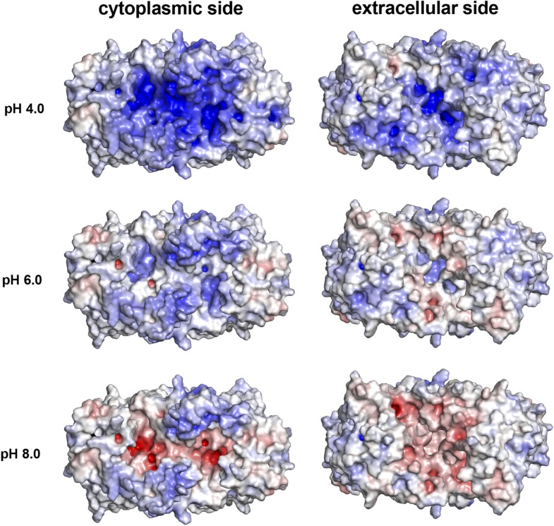
Figure 2—figure supplement 1. pH-dependent charge distribution.

Figure 2—figure supplement 1.

The surface potential of the PaNhaP dimer was calculated at pH 4, pH 6, and pH 8 by APBS and visualized in PyMOL. At pH 4 the cytoplasmic surface is strongly positively charged. At pH 6, the extracellular surface and the cytoplasmic funnel are largely neutral. At pH 8 the extracellular surface is predominantly negatively charged and the cytoplasmic funnel is strongly negatively charged, inhibiting the release of substrate ions. The ion-binding site is located at the bottom of the cytoplasmic funnel.