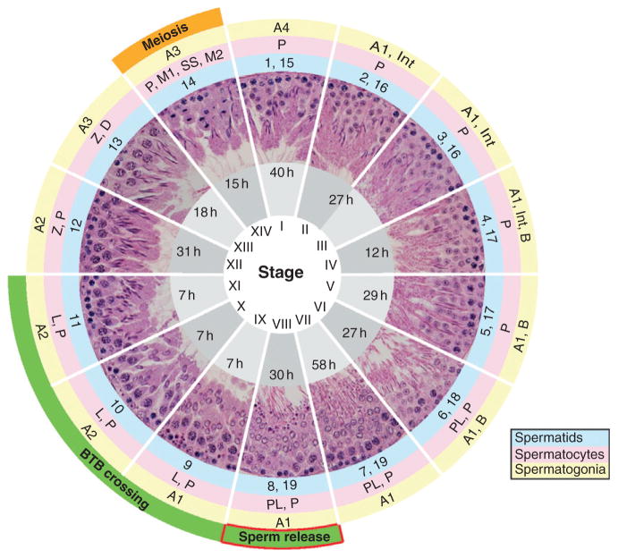
Figure 5.1.
Seminiferous epithelial cycle in the rat testis. These 14 images represent stages of the seminiferous epithelial cycle obtained from paraffin-embedded cross-sections of the adult rat testis stained with hematoxylin and eosin. Stages are noted as roman numerals. Annotations in gray shaded areas indicate the approximate duration of each stage in hours (h). Germ cells are divided into spermatogonia (outer yellow circle), spermatocytes (middle pink circle), or spermatids (inner blue circle). Spermatogonia include types A1–A4, intermediate (Int) and B (yellow circle). Spermatocytes (i.e., preleptotene (PL), leptotene (L), zygotene (Z), pachytene (P), and diplotene (D)), primary spermatocytes in meiosis 1 (M1), secondary spermatocytes (SS), and secondary spermatocytes in meiosis 2 (M2) are also shown (pink circle). Finally, spermatid differentiation spans steps 1–19 (blue circle). Important cellular events are noted in the outermost layer as orange and green shaded areas. Spermiation takes place at stage VIII, concurrent with the transit of preleptotene spermatocytes across the BTB during stages VIII–XI. M1 and M2 take place at stage XIV.

