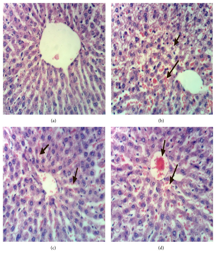
Figure 2.
(a) Liver of rat from group 1 showing the normal histological structure of hepatic lobule, (b) liver of rat from group 2 showing steatosis of hepatocytes and focal hepatic necrosis associated with inflammatory cells infiltration, (c) liver of rat from group 3 showing slight congestion of hepatic sinusoids, and (d) liver of rat from group 4 showing congestion of central vein and hepatic sinusoids (H&E ×400).

