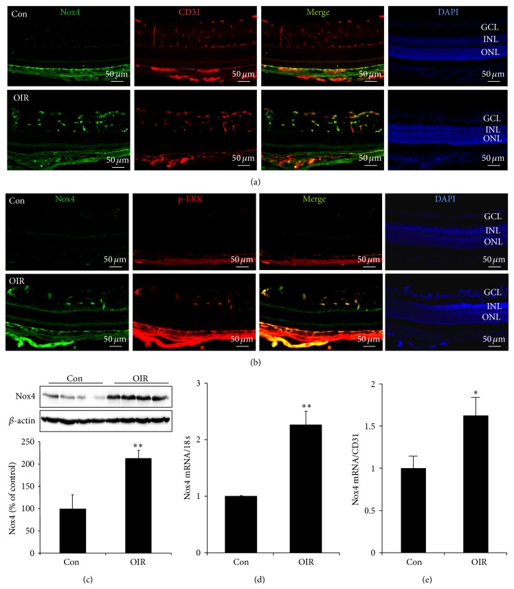
Figure 2.
Nox4 expression in retinas of OIR mice at retinal NV phase. (a) Immunostaining of Nox4 (green) in retinal cryosections from P17 OIR and control mice. CD31 (red) was used to label retinal vessels. (b) Expression of phosphorylated ERK (red) and its colocalization with Nox4 (green) in the retina in P17 OIR and control mice were determined by immunostaining. (c) Expression of Nox4 protein in the retina in P15 OIR and control mice was determined by Western blot analysis and semiquantified by densitometry. n = 4 for each group. ** P < 0.01 versus Con. (d-e) mRNA of Nox4 in the retina in P15 OIR and control mice was measured by real-time RT-PCR and normalized to 18 s (d) or to CD31 (e). n = 4 for control group and n = 6 for OIR group. ** P < 0.01 versus Con.

