
Figure 2. Negative-stain EM and AFM of disulphide-locked suilysin.
(A) Negative-stain EM disulphide-locked suilysin (ds-SLY) on egg PC:cholesterol monolayers (45:55%), locked in the prepore state (−DTT). (B) as (A), for disulphide-locked suilysin incubated in the presence of 5 mM DTT in solution to reduce the disulphide bridge, so that the suilysin is rapidly converted to the pore conformation. (C) AFM of densely packed suilysin prepores, confined to the egg PC-rich domain of a phase-separated egg PC:DDAB:Cholesterol (33:33:33%) supported lipid bilayer, with its corresponding height distribution (D) referenced to the membrane surface.
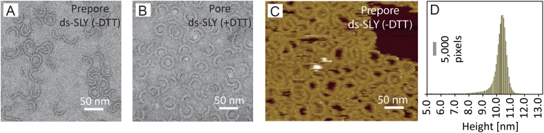
Figure 2—figure supplement 1. Radius of curvature for arc-shaped suilysin assemblies in the prepore and pore states.

(A) For wild-type suilysin (WT-SLY), the curvature
distribution of the arc-shaped assemblies shows a sharp peak close to the
radius of the complete ring with 37-fold symmetry. (B) For
disulphide-locked suilysin in the prepore state (ds-SLY, −DTT),
the curvature distribution peaks at slightly lower radius but also shows
a larger spread to radii of curvature far exceeding that of the complete
37-mer ring. (C) When the disulphide bridge is unlocked by
DTT (ds-SLY, +DTT, cf. Figure
2B), the insertion of the transmembrane hairpins in the lipids
and formation of the β-barrel leads to a shaper distribution of
the arc-shaped oligomers, similar to the wild-type suilysin.
Corresponding negative stain EM views are shown under each plot. These
observations are further evidence that the prepore intermediate is a
structurally flexible state. The arrows refer to the circular fit radius
to a 37-mer suilysin ring, which corresponds to 13.9 nm for the prepore
assembly and 15.1 nm for the pore state assembly. Scale bars: 25 nm.