. 2014 Dec 2;3:e04247. doi: 10.7554/eLife.04247

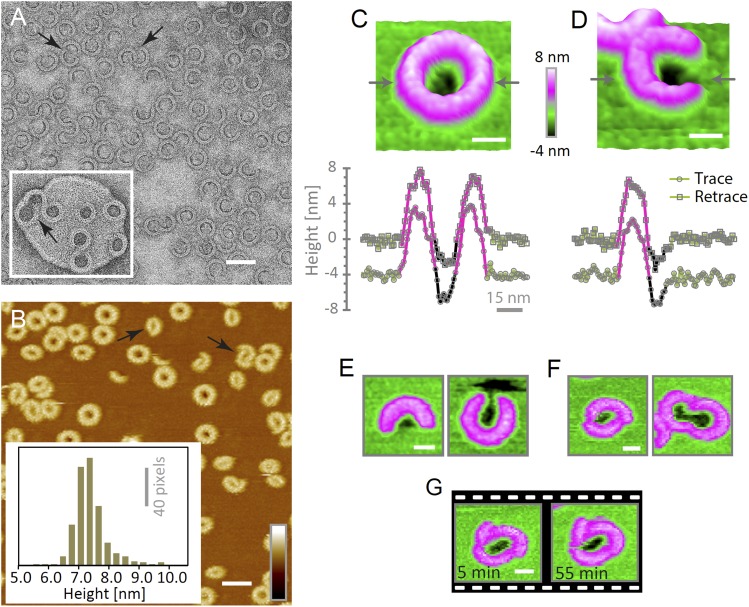
Figure 4. Suilysin assembles into ring- and arc-shaped oligomers that perforate the membrane.

(A) Negatively stained EM of arc- and ring-shaped assemblies of wild-type suilysin on an egg PC:cholesterol (45:55%) lipid monolayer, and (inset) on a liposome of egg PC:cholesterol (45:55%). (B) AFM topography of wild-type suilysin on a supported egg PC:cholesterol (67:33%) lipid bilayer. The wild-type suilysin extends 7–8 nm above the lipid bilayer background, as indicated by the height histogram for 402 individual particles (inset). (C) The AFM topography of a complete suilysin ring reveals a circular hole (dark) in its lumen, whereas the lipid bilayer surrounding the ring remains intact (green). (D) The topography of a suilysin arc shows a hole (dark) in the membrane only partially enclosed by the suilysin assembly. Images in C and D are shown in a 15° tilted representation, and height profiles measured across the ring/arc confirm membrane perforation. (E) Examples of wild-type suilysin arcs of different lengths. Transmembrane holes are consistently observed. (F) Examples of interlocked-arc assemblies. As shown in the right image, the membrane area removed by the two arcs is larger than the hole in the complete ring (C). (G) Sequence of AFM images of the same interlocked-arc assembly, stable for at least 50 min. Scale bars AB: 50 nm, CG: 15 nm, full z colour scale BG: 12 nm.

DOI: http://dx.doi.org/10.7554/eLife.04247.013

Figure 4.

Figure 4—figure supplement 1. Suilysin pore assemblies by EM and AFM.

Figure 4—figure supplement 1.

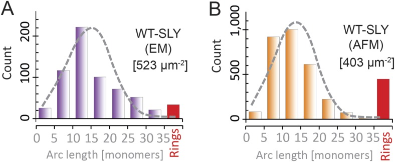
The arc-length distributions for wild-type suilysin as measured by negative-stain EM on monolayers (A) and the corresponding AFM data on supported lipid bilayers (B). The grey dashed curves denote the fits of the experimental data with the oligomerization model with ka/kb = 3.461 ± 0.019 µm2 (A), and 3.468 ± 0.004 µm2 (B). The numbers in brackets in (A) and (B) denote the number of monomers per square micron. When using similar lipid compositions (egg PC:Cholesterol 67:33%) and incubation conditions (27°C), both experiments yield very similar distributions.
Figure 4—figure supplement 2. Suilysin is a monomer in solution.

Figure 4—figure supplement 2.

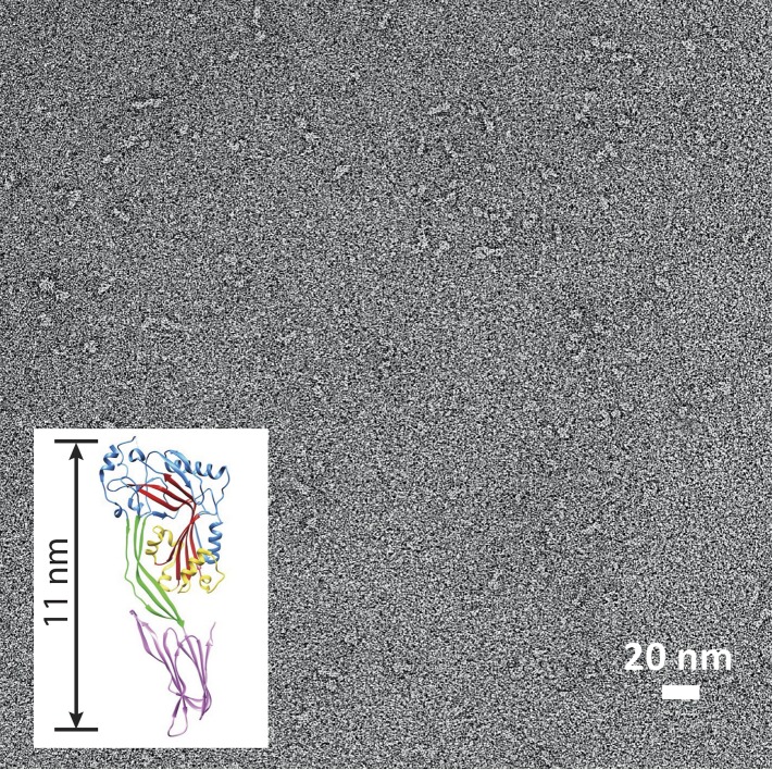
Negative-stain EM of a carbon grid after incubation with wild-type suilysin at a concentration of 10 µg/ml. In the absence of lipids, only monomers are observed. Inset: crystal structure of suilysin (Xu et al., 2010), for comparison.
Figure 4—figure supplement 3. AFM assays of wild-type suilysin (WT-SLY) doped with disulphide-locked suilysin (ds-SLY).

Figure 4—figure supplement 3.

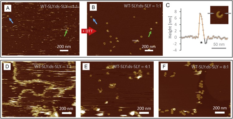
AFM topographic images of WT-SLY doped with an equimolar amount of ds-SLY (AC) in solution and incubated on egg PC: cholesterol (67:33%) lipid bilayers. (A) The presence of ds-SLY largely traps the WT-SLY in the prepore conformation with only few pores observed (blue and green arrows). Addition of ∼5 mM DTT unlocks the disulphide bond and AFM imaging in the same area (B) reveals more arcs and rings of suilysin in the pore state. (C) Line profile from a suilysin arc after addition of DTT, demonstrating that the membrane is perforated (*). (DF) AFM images of WT-SLY doped with decreasing amounts of ds-SLY in solution and incubated on DOPC:sphingomyelin:cholesterol, 33:33:33%. (D) At 1:1 ratios of WT-SLY:ds-SLY, the result is similar to (A) with very few suilysin pores observed and prepore locked oligomers prevalent and confined by and at the lipid boundaries. (E) At lower amounts of dopant (WT-SLY:ds-SLY = 4:1), more suilysin pores become visible, with some remaining prepore suilysin oligomers observed as higher arcs and rings and as diffuse streaks. This demonstrates that on reducing the proportion of ds-SLY, the WT-SLY recovers its effectiveness in forming pores in the membrane. (F) With the relative ds-SLY proportion reduced even further (WT-SLY:ds-SLY = 8:1), mostly suilysin pores are prevalent.