Figure 3.
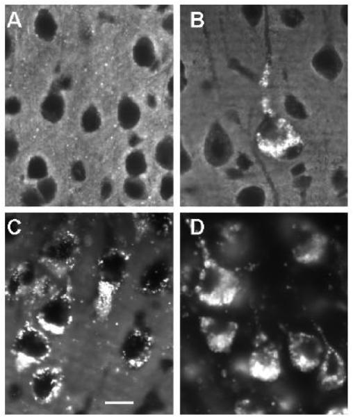
Filipin histochemical staining for unesterified cholesterol in cerebrocortical neurons in multiple lysosomal diseases. A: Wt. (12 weeks old) B: MPS IIIA disease (12 weeks old). C: Niemann-Pick disease type C (8 weeks old). D: GM1 gangliosidosis (12 weeks old). Note that neurons in Wt brain exhibit no significant filipin labeling of somata, whereas in each of the lysosomal diseases there is substantial filipin labeling of individual neurons. In MPS IIIA disease, some neurons are clearly more affected than others, whereas in Niemann-Pick C and GM1 gangliosidosis all neurons are positive. Filipin staining in GM1 was substantial and appeared to exceed that of Niemann-Pick C in late stage disease. Calibration bar in C equals 12 µm and applies to all.

