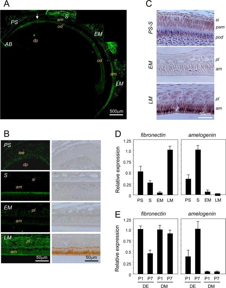
Fig 1. Analysis of fibronectin expression during tooth development.
A. Immunofluorescence analysis of fibronectin expression using an anti-fibronectin antibody in sagittal sections from 6-week-old mouse incisors. The incisors were separated into the apical bud (AB), presecretory (PS), secretory (S), early maturation (EM), and late maturation (LM) stages. B. Immunofluorescence and immunohistochemical results are shown at a higher magnification. Fibronectin was expressed in the basal lamina at the S stage and in the papillary layer at the LM stage. C. Fibronectin mRNA was detected using in situ hybridization in the basal lamina, pre-odontoblasts, pre-ameloblasts from PS to S (PS-S) and LM ameloblasts, with lower expression seen during the EM stage. D. The expression levels of fibronectin and amelogenin were investigated by real-time PCR using PS, S, EM, and LM dental epithelial cells separated from the incisor. E. The expression of fibronectin in dental epithelial (DE) and dental mesenchymal (DM) cells isolated from the molars of P1 and P7 mice (n = 5) was investigated by real-time PCR. Amelogenin mRNA expression increased in DE cells, while fibronectin mRNA expression decreased with differentiation. iee, inner enamel epithelium; dp, dental pulp; si, stratum intermedia; am, ameloblasts; pam, pre-ameloblasts; od, odontoblasts; pod, pre-odontoblasts; pl, papillary layer.

