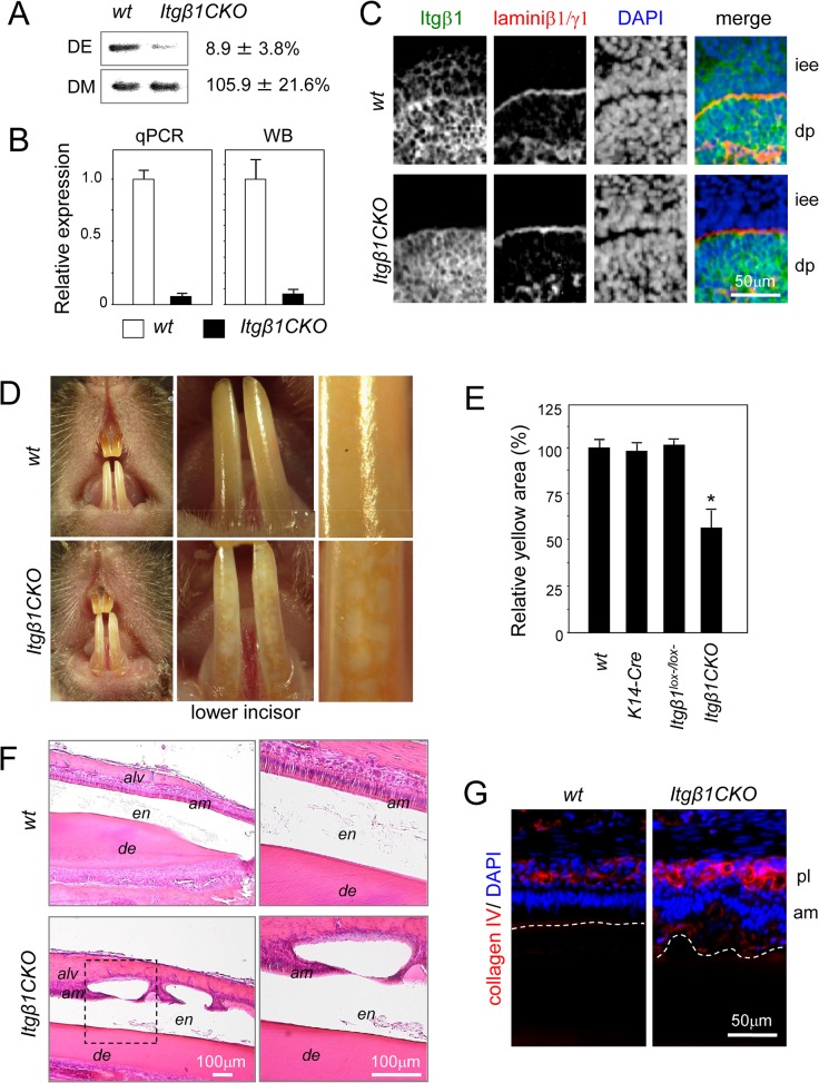
Fig 4. Phenotype of incisors from Itgβ1CKO mice.
Itgβ1 was deleted from cells that expressed cytokeratin 14. A. Southern blot analysis of genomic DNA from P1 molars using a probe for exon 3. Most of the region containing exon 3 was removed by Cre recombinase under the control of the K4 promoter in the dental epithelium. B. Expression levels of β1 integrin mRNA and protein in P1 molar tooth germs. C. Immunohistolocalization of β1 integrin in E16.5 molar tooth germs. D. Frontal view of a 8-week-old wild type (WT) and homozygous Itgβ1 conditional knockout (CKO) mouse. Many vitiligo patches are visible in the image of the lower incisor from an Itgβ1CKO mouse. E. The lower incisors were observed under higher magnification and the total size of the yellow areas was measured. Yellow areas were markedly decreased in the incisors from Itgβ1CKO mice (n = 5). F. Sagittal sections of lower incisors from a 6-week-old WT and Itgβ1CKO mouse were stained with hematoxylin and eosin (H-E). High magnifications of the areas enclosed by the dashed box in the left panel. Cysts were formed in the late maturation (LM) stage. G. Immunostaining using an anti-collagen IV antibody. Nuclei were stained using DAPI. The image shows an irregular and multilayer arrangement of ameloblasts in the incisor from an Itgβ1CKO mouse. The dotted line shows the border of enamel and ameloblasts. *P < 0.01. iee, inner enamel epithelium; dp, dental pulp; alv, alveolar bone; en, enamel; de, dentin; am, ameloblasts; pl, papillary layer.

