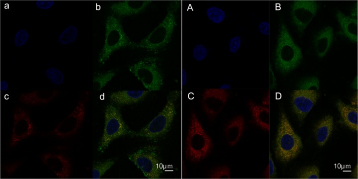
Fig 8. Confocal microscopy images showing the intracellular localization of coumarin-6-loaded PEG-PLA micelles in A549 cells.
(a) nucleus, (b) PEG-PLA micelles, and (c) lysosomes were distinguished using Hoechst 33342 (blue), coumarin 6 (green), and Lyso Tracker Red (red). (d) Yellow represents the colocalization of Lyso Tracker Red with Green (coumarin-6-loaded PEG-PLA micelles). A, B, C, and D show the localization of coumarin-6-loaded mixed micelles.

