
FIGURE 2.
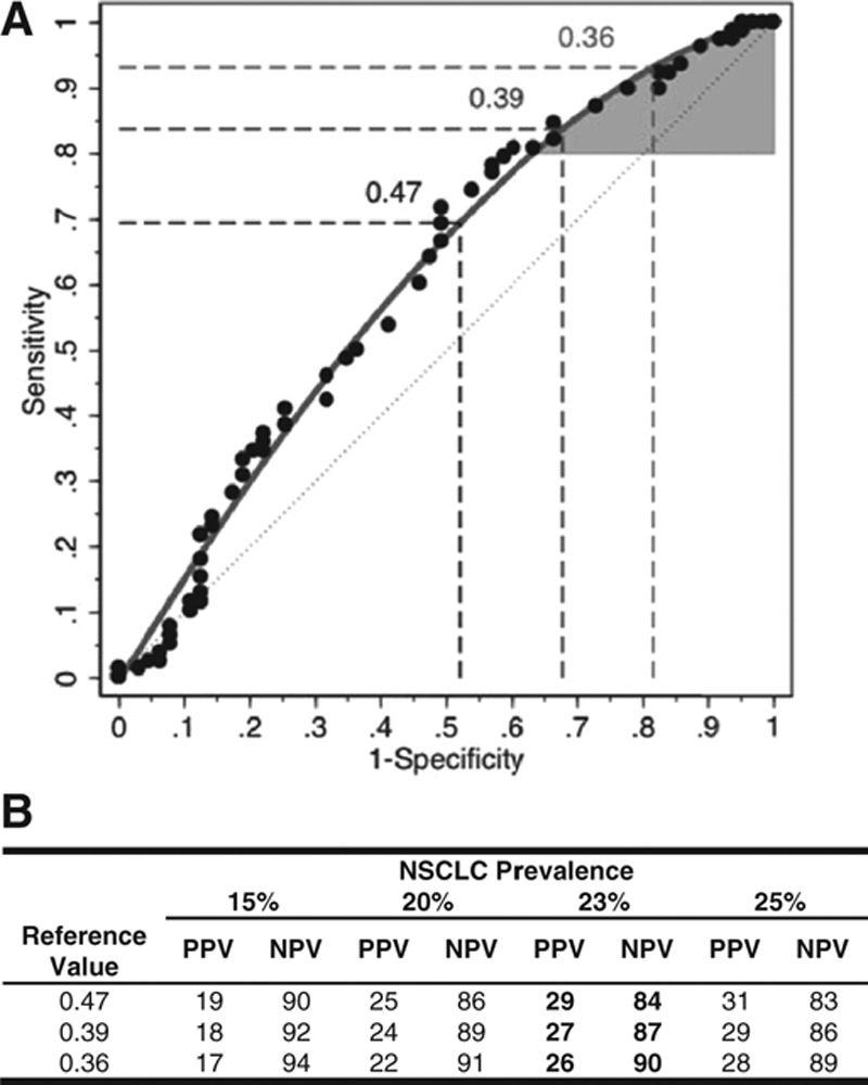
Protein expression classifier validation. The protein expression classifier yields a score between 0 and 1, with lower values associated with a higher probability of a benign etiology, based on the identification and quantification of specific plasma proteins (Supplemental Materials, Supplemental Digital Content, http://links.lww.com/JTO/A773). A, The statistical performance of the classifier was validated with a lower 95% confidence bound for the partial area under the curve (pAUC) of 0.026, which was greater than the targeted pAUC of 0.02; the performance of the classifier was validated at predefined reference values from 0.38 to 0.47. Shown are the receiver operating characteristic (ROC) curves based on the raw (circles) and fitted (grey line) data, along with the ROC curve associated with chance (grey dashed line). The maximal classifier score in the study for use as a reference value to assign a likely benign classification was 0.47, using the fixed-sequence procedure.27,28 Therefore, the region bounded by the grey ROC curve and a sensitivity of 80% represents the classifier’s partial area under the ROC curve (AUC) (shaded in grey). The associated sensitivity and specificity values (%) for the reference value of 0.47 are indicated (dashed lines). Similar data for reference values of 0.39 and 0.36, which correspond to NPVs of 87% and 90%, respectively, are indicated (dashed lines). B, A reference value is a specific score at or below which the classifier may assign a likely benign result to a given plasma specimen, based on the measured values of the classifier’s constituent proteins. Classifier scores between 0.18 and 0.47 may be used as a reference value. Shown are the positive and negative predictive values (PPV and NPV, respectively) for classifier reference values of 0.36, 0.39, and 0.47 using cancer prevalences of 15%, 20%, 23%, and 25%. The value of 0.47 is shown based on the prespecified criterion for validation of the classifier using the fixed sequence procedure; and the values of 0.36 and 0.39 are shown to illustrate a diagnostic performance of 90% NPV and an intermediate value, respectively.