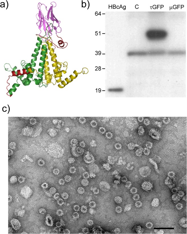
Fig 7. τGFP expressed in plants forms VLPs.

a) Predicted structure of the τGFP tandibody protein (Swiss-Prot model): green: core 1, yellow: core 2, pink: anti-GFP nanobody, red: linkers. b) Western blot of crude plant extracts. C: empty vector control, τGFP: tandem HBcAg construct with anti-GFP VHH in the core 2 MIR, μGFP: monomeric HBcAg containing anti-GFP VHH in the MIR. The 39 kDa band found in all plant extracts is non-specific. c) Electron micrograph of plant-produced τGFP particles purified by sucrose cushion. Scale bar 100 nm.
