
Fig 8. Plant-produced τGFP particles bind GFP.
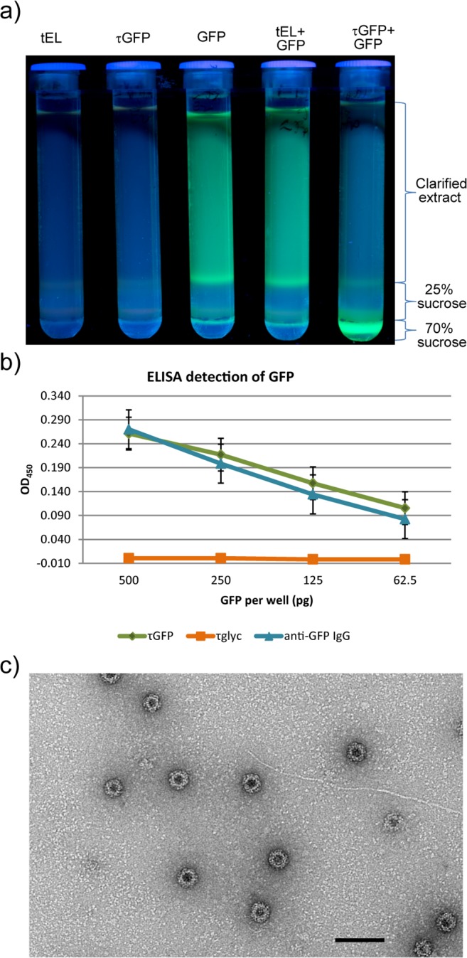
a) Ultracentrifuge tubes containing sucrose cushions photographed under UV light after ultracentrifugation. GFP-associated fluorescence remains in the supernatant when GFP-containing plant lysate is centrifuged alone or mixed with tEL-containing plant lysate; but migrates through the cushion when GFP-containing and τGFP-containing plant lysates are mixed. b) Detection of GFP by sandwich ELISA, after coating wells with τGFP (green), τglyc (orange) or an anti-GFP polyclonal IgG (blue) and adding GFP to the wells at four different concentrations after blocking. Detection is horseradish peroxidase—mediated ECL, and signal is net of background. Error bars are standard error. c) Electron micrograph of plant-produced τGFP particles in the presence of GFP, purified by sucrose cushion and size exclusion chromatography. Scale bar 100 nm.