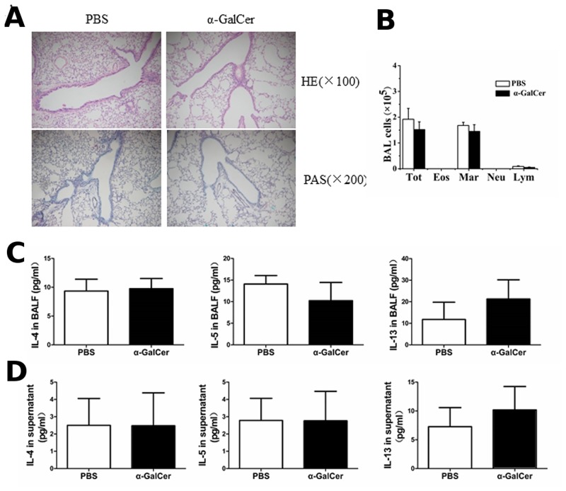
Fig 2. α-GalCer administration cannot induce the Th2 inflammatory response in WT mice.
A. Histopathological analysis using the hematoxylin-eosin (H&E) staining and periodic acid-Schiff (PAS) staining of lung tissue sections from mice treated with α-GalCer or PBS. B. Total cell and differential cell counts in the BALF were obtained using a standard hemocytometer. N = 5 per group. Tot, total cell counts; Mar, macrophages; Eos, eosinophils, Neu, neutrophils; and Lym, lymphocytes. C. BALF were collected 3 days after α-GalCer or PBS administration and cytokine (IL-4, IL-5, and IL-13) production was analyzed by ELISA. N = 4–5 per group. D. Splenocytes were obtained from mice with α-GalCer or PBS administration and re-stimulated with 500 μg OVA in vitro. After 72 h, culture supernatants were collected and cytokine production was analyzed by ELISA. N = 5 per group.

