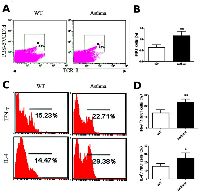
Fig 3. iNKT cells are activated in the OVA-induced asthma model.
A. The percentage of iNKT cells (PBS-57/CD1d+ TCR-β+) in lung MNCs. The gating used for iNKT cells (gate D) and the corresponding percentages are shown in each dot plot. B. Percentage of iNKT cells in lung MNCs from OVA-induced asthmatic mice and WT mice. N = 5 per group and **P < 0.01. C. Lung iNKT cells producing IFN-γ and IL-4. PBS-57/CD1d and TCR-β double-positive cells were examined for IFN-γ (top) and IL-4 (bottom) secretion. Numbers in each dot plot indicate the percentage of positive cells. D. Percentage of IFN-γ- and IL-4-expressing lung iNKT cells. N = 5 per group. *P < 0.05, **P < 0.01.

