Fig. 3.
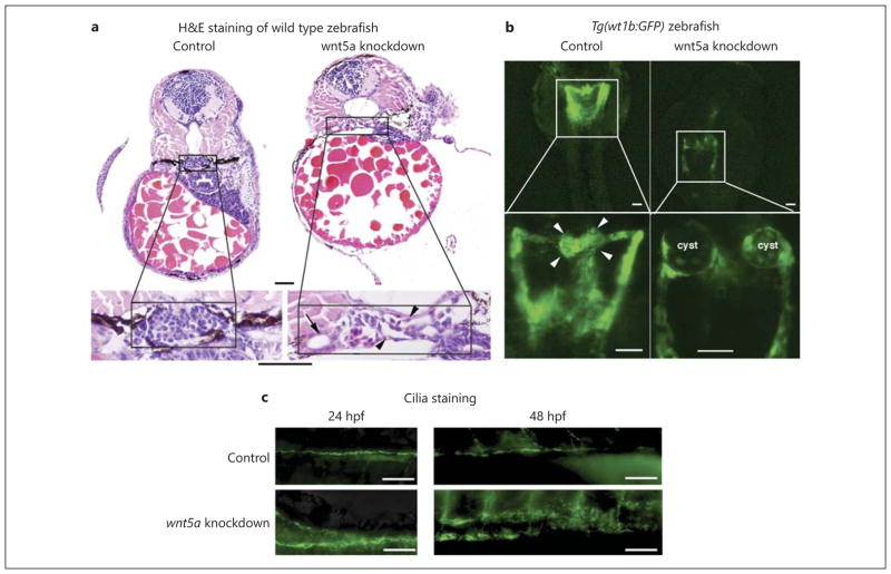
wnt5a knockdown by antisense morpholinos results in pronephric cyst formation. a HE staining of the transverse histological sections of 72-hpf morphant embryos reveals disrupted glomerular structures and dilated glomeruli (arrowhead) and renal tubules (arrow) after wnt5a knockdown by both AUG- and splice-MOs (image is from control and AUG-injected zebrafish; splice-MO injection showed similar results, but image is not shown here). Bar = 20 μm. b Representative images of the pronephros of 48-hpf Tg(wt1b:GFP) transgenic zebrafish embryos injected with phenolred control and wnt5a AUG-MO. Cystic glomeruli are clearly visible in the wnt5a morpholino-injected animals but not in the control (phenol-red) ones. Arrowheads indicate glomeruli. (Splice- MO injection showed similar results, which is not shown here.) Bar = 50 μm. c Representative images of the ciliary staining in pronephros at 24 and 48 hpf. wnt5a knockdown results in disordered ciliary structure compared with injection control. Bar = 50 μm.

