Fig. 5.
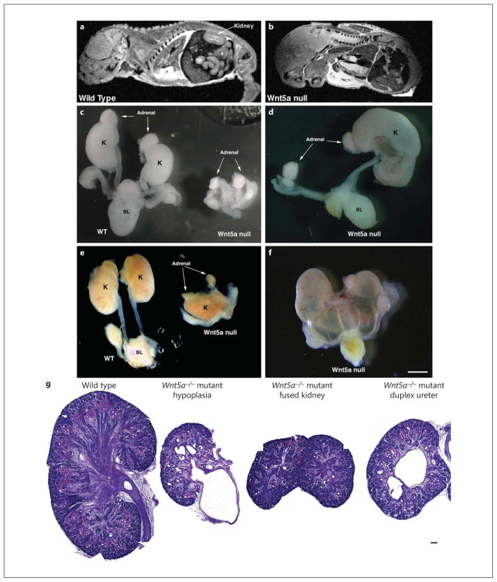
Metanephric kidney development is severely compromised in Wnt5a global knockout mice as determined by MRI, fine dissection, and HE staining. a, b MRI of E16.5 mouse embryos. A normal-sized kidney is seen in the wild-type mouse embryo (a), whereas a Wnt5a−/− mouse embryo shows absence of kidney formation (b). Due to the skeletal abnormalities, it was difficult to identify identical sections in control and Wnt5a−/− mice. c–f Wild-type and Wnt5a−/− mouse kidneys (when tissue was present) and urinary tracts were dissected at E16.5. The kidneys from Wnt5a−/− mutants shows pleiotropic, but severe, kidney phenotypes, including bilateral kidney agenesis (c), unilateral kidney agenesis (d), fused kidneys (e) and hydroureteronephrosis (f). c–f Bar = 1 mm. g HE staining of the embryonic kidneys from wild-type and Wnt5a−/− mice. Histologic sections of E16.5 wild-type (left) and Wnt5a−/− (right) embryonic kidneys shows markedly reduced kidney tubules and glomeruli in Wnt5a−/− embryos. There is also significant dilatation of the renal tubules and hydronephrosis in the Wnt5a−/− mutant embryos. g Bar = 50 μm.

