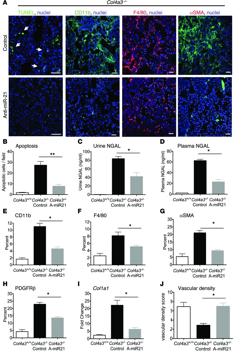
Figure 4. Tubular injury and myofibroblast and leukocyte expansion are all attenuated by anti–miR-21.
(A) Images of apoptotic tubular epithelial cells (arrows), macrophages, and myofibroblasts. (B–D) Quantification of tubular apoptosis and the tubular injury marker NGAL in urine and plasma at 9 weeks. (E–H) Quantification of kidney macrophages (CD11b, F4/80), fibroblasts/PCs (PDGFRβ), and myofibroblasts (αSMA) at 9 weeks. (I) qPCR for fibrogenic transcripts. (J) Quantification of peritubular capillary density. *P < 0.05; **P < 0.01, Mann-Whitney U test. n = 12/group. Scale bars: 50 μm.

