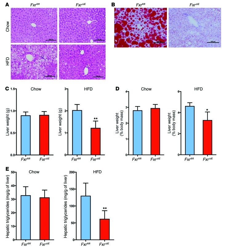
Figure 3. Intestine-specific Fxr disruption protects against HFD-induced NAFLD.
(A) Representative H&E staining of liver sections from Fxrfl/fl and FxrΔIE mice after 14 weeks of chow or HFD feeding. Scale bars: 100 μm. n = 4–5 mice per group. (B) Representative Oil red O staining of liver sections from Fxrfl/fl and FxrΔIE mice after 14 weeks of HFD feeding. Lipids stained positive (red color) with Oil Red O. Scale bars: 100 μm. n = 5 mice per group. (C) Liver weights of Fxrfl/fl and FxrΔIE mice after 14 weeks of chow or HFD feeding. n = 4–5 mice per group. (D) Liver weight/body weight ratios of Fxrfl/fl and FxrΔIE mice after 14 weeks of chow or HFD feeding. n = 4–5 mice per group. (E) Liver triglyceride contents in Fxrfl/fl and FxrΔIE mice after 14 weeks of chow or HFD feeding. n = 4–5 mice per group. All data are presented as the mean ± SD. *P < 0.05 and **P < 0.01 (2-tailed Student’s t test) compared with Fxrfl/fl mice.

