
Figure 1. Molecular mechanism of autophagosome formation and disposal of cellular material.
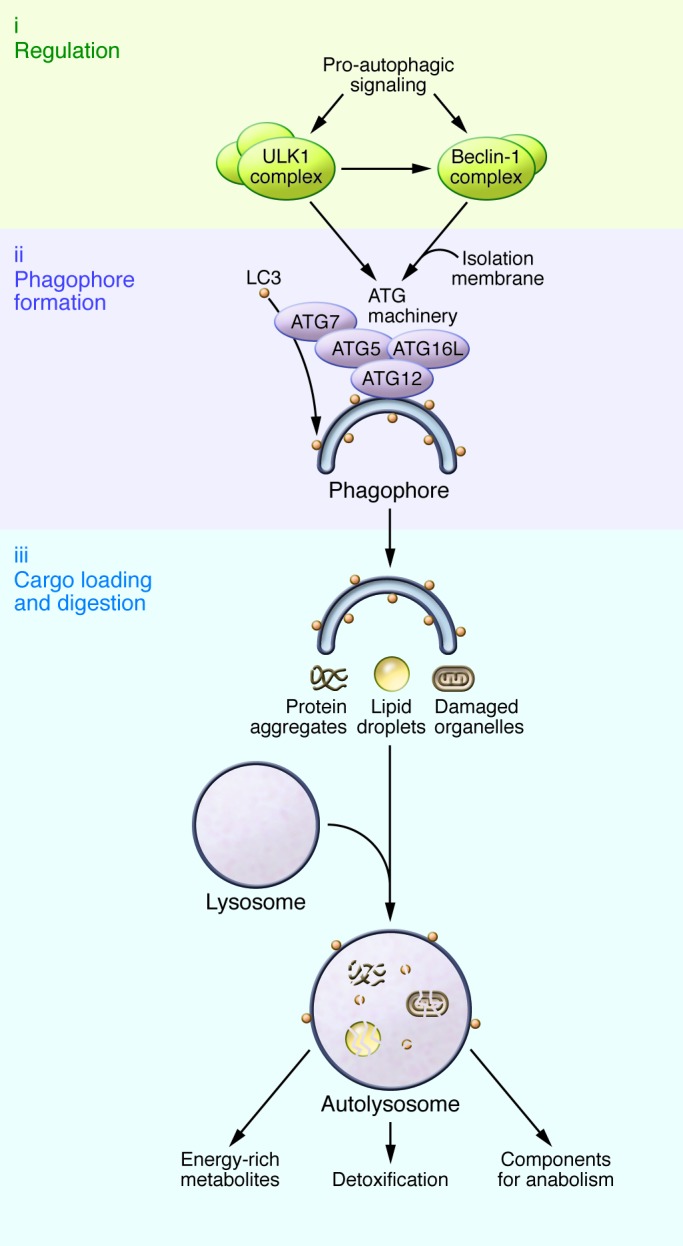
Pro-autophagic signals such as nutrient or growth factor depletion activate regulatory components of the autophagic machinery, such as the ULK1 and Beclin-1 complexes. Isolation membranes form at ER-mitochondrial interfaces or recycling endosomes under the guidance of the ATG machinery, which, among other components, consists of ATG7, ATG5-ATG12, and ATG16L. ATG7 drives the lipidation of LC3, which incorporates into the incipient phagophore to mediate the recognition and loading of cargo, such as protein aggregates, damaged organelles, or lipid droplets. The mature autophagosomes (not shown) fuse with lysosomes to form autolysosomes, through which the engulfed cellular material gets digested. As a result, potentially harmful misfolded protein aggregates or damaged organelles are detoxified, and their degradation products can be used to replenish cellular energy reserves and anabolic reactions.