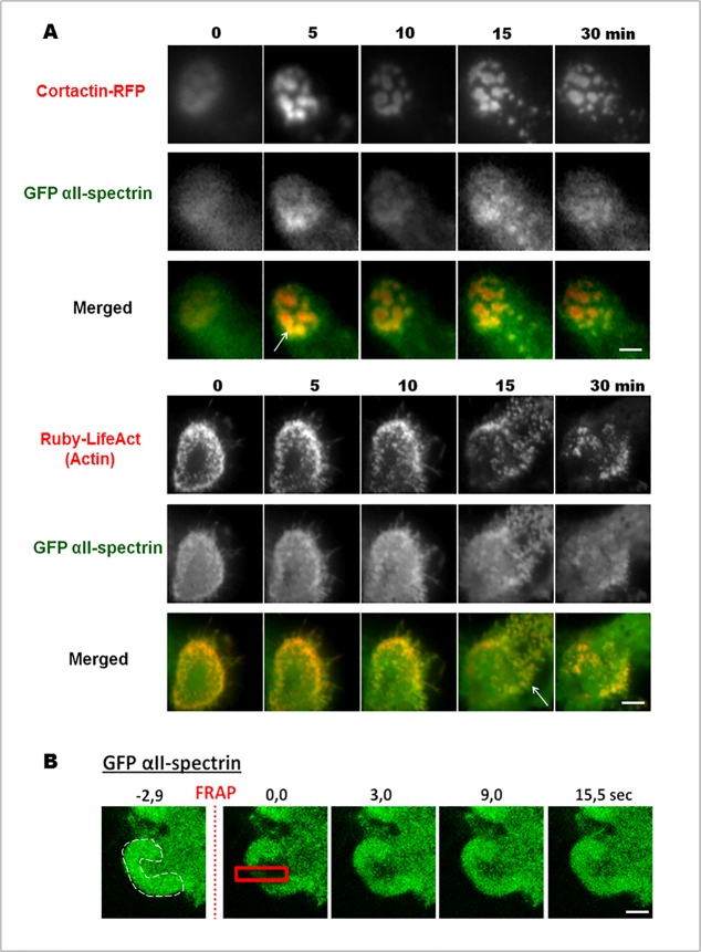
Fig 3. GFP αII-spectrin dynamics in invadosomes.
(A) Extracted images from time series (min) from representative observations by TIRF microscopy of living SrcY527F-MEF cells expressing full length of αII-spectrin fused to GFP an invadosome marker fused to RFP, cortactin, or the actin marker Ruby-LifeAct. Ten observation fields were analyzed from at least three different experiments. GFP-αII-spectrin accumulated in isolated or invadosome rings, during their expansion or disorganization. These data link αII-spectrin with the intense actin remodeling associated with invadosome dynamics. (B) To confirm this point, net flux of GFP- αII-spectrin was analyzed by FRAP technology. After a 2.9 sec photobleaching in the red square, αII-spectrin fluorescence starts to reappear after only 3.0 sec, and total recovery of the fluorescence in the photobleached area occurred at 15.0 sec. The invadosome ring is identified by a white dash line here in a portion of cell in the first time. Relevance of the results were obtained by analyzing 10 observation fields from at three different experiments. Scale bar: 3 μm.

