
Fig 7. Localization of different PROVIR-GFP proteins in N. benthamiana leaves and protein co-localization with the plasma membrane marker protein PIP-mCherry by confocal microscopy.
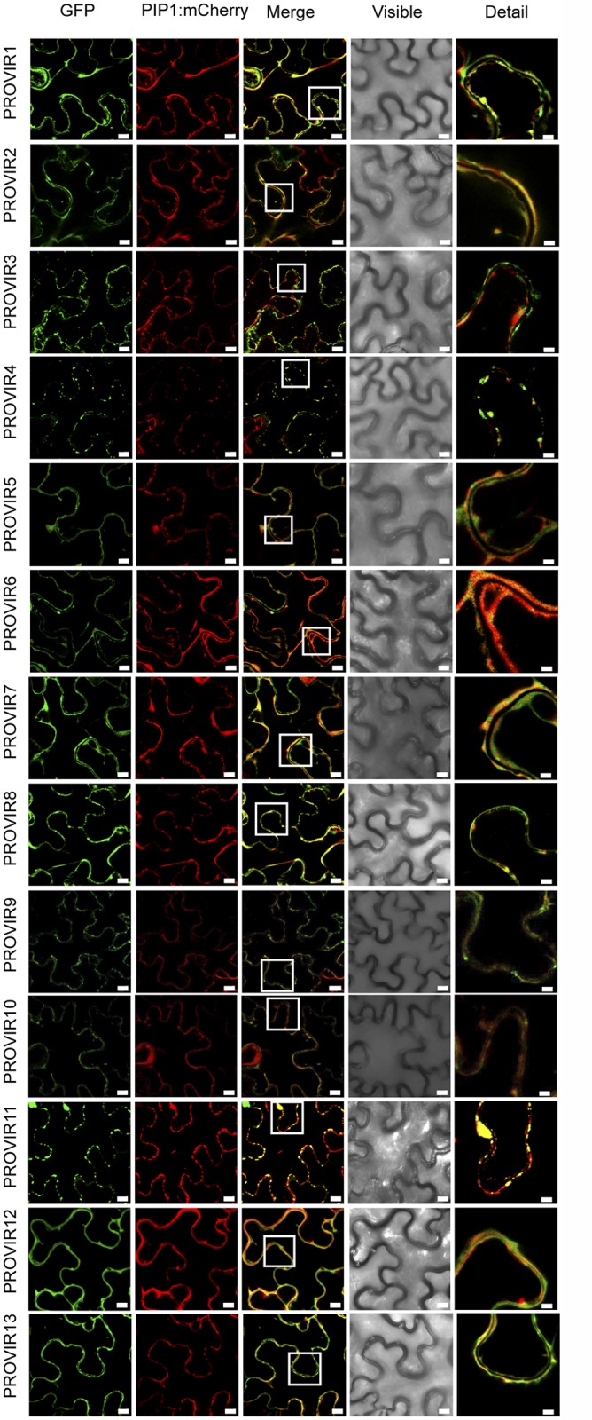
Expression of PROVIR1-to-13-GFP (72 h.p.i.) resulted in distinct pericellular fluorescence patterns distributed either linearly along the plasma membrane or forming punctuated foci resembling small membrane clusters. Co-expression of PROVIR-GFP with the plasma membrane marker PIP-mCherry facilitated tracing the plasma membrane. Scale bars are 8 μm, except for the right longitudinal panel where scale bars are 2 μm. This provides a magnified detail of the boxed sector in the intermediate panel merging GFP and mCherry-derived fluorescence.