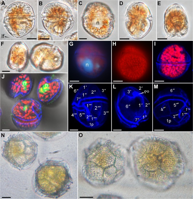
Fig 1. Light microscopy pictures of Fukuyoa paulensis gen. et sp. nov. (A-M) and Goniodoma polyedricum (N-O).
(A-F) Live cells observed under a Zeiss Imager.A2 with Nomarski differential interference contrast (D.I.C.) optics at ×1000 magnification. (A) Left lateral view. Note the longitudinal flagellum. (B-C) Right lateral view. Note the nucleus. (D) Dorsal view. (E) Antapical view. Apical and ventral views. (G-H) Glutaraldehyde-fixed cells observed under an Olympus BX51 epifluorescence microscope at ×1000 magnification. (G) See the nucleus stained with DAPI. (H) Epifluorescence image of chloroplasts. (I-M) Glutaraldehyde-fixed cells observed under an inverted Leica TCS SP8 AOBS confocal microscope at ×630 magnification. The plates were stained with Fluorescent Brightener 28, and the nuclei were stained with SYBR Green I. (I) Overlay image of the cell showing the plastids. (J) See the nucleus stained with SYBR Green I. (K-M) Overlay image showing the thecal plates stained with Fluorescent Brightener 28. (N-O) Live cells of Goniodoma polyedricum isolated for PCR. Pictures taken at ×600 magnification with a Nikon TS-100 inverted microscope equipped with a SONY CyberShot DSC-W310 camera. n = nucleus; lf = longitudinal flagellum. Scale bar 10 μm.

