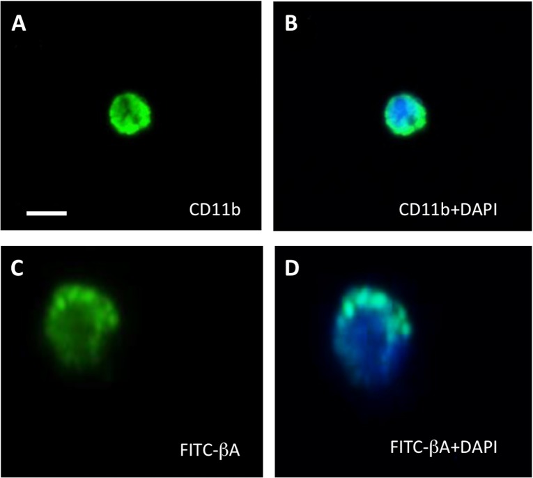
Fig 3. Microscopy analysis of CD11b immunostaining and phagocytosis of FITC-beta-amyloid (Aβ).
Following 24 h incubation in the absence (A&B) or presence (C&D) of FITC-Aβ, CD11b-positive cells (isolated from adult C57BL/6N mice) were evaluated for CD11b surface marker staining (A) and engulfment of FITC-Aβ1–42 (C). Images illustrate apparent fluorescent staining (green) in healthy cells stained positive for nuclear (blue) DAPI (B&D). Cells displayed diffuse cytoplasmic granulated/ punctate staining of phagocytosed FITC-Aβ1–42: (C&D). Scale bar = 10 μm (A&B), 5 μm (C&D).

