
Fig 6. Protein levels of APP, catalase, and cytokines in monocyte-infused APPSwDI mice.
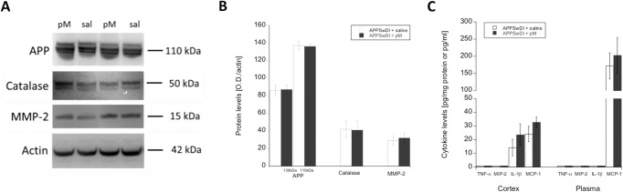
Following monocyte infusions, brains were collected and cortical extracts were evaluated for amyloid precursor protein (APP), catalase, or matrix metalloproteinase-2 (MMP-2) by Western blot (A&B; n = 4–6 per animal group). In addition, cortex (n = 6 per animal group) and plasma (n = 4 per animal group) were evaluated for tumor necrosis factor-α (TNF-α), macrophage inflammatory protein-2 (MIP-2), interleukin-1β (IL-1β), and monocyte chemotactic protein-1 (MCP-1) levels by ELISA (C). APPSwDI mice receiving young peripheral blood monocyte (pM) i.v. infusions are indicated with a shaded gray bar. APPSwDI mice receiving saline (sal) served as negative controls and are indicated with a white bar. Bar graphs display the mean ± SEM (error bars) of protein levels. Statistical analysis was performed using a Student’s t-test (all not significant).