
Figure 2. Color M-Mode Echocardiography.
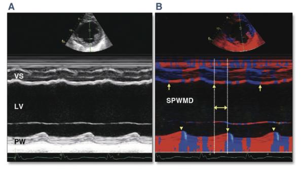
(A) Conventional M-mode echocardiography. Evaluating earliest activation is difficult because of hypokinesis and wall thinning from prior infarction. (B) M-mode echocardiography with color-coded tissue velocity. Activation may be more easily assessed visually as a color change from blue to red (arrows demonstrate change in color denoting myocardial contraction for the ventricular septum [VS], arrow-heads for the posterior wall [PW]). LV = left ventricular; SPWMD = septal to posterior wall motion delay. Reprinted, with permission, from Anderson LJ, Miyazaki C, Sutherland GR, Oh JK. Patient selection and echocardiographic assessment of dyssynchrony in cardiac resynchronization therapy. Circulation 2008;117:2009-23.