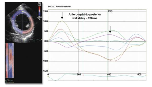
Figure 4. Two-Dimensional Speckle Tracking.

Apical view of the left ventricle demonstrating 2-dimensional speckle tracking for radial strain assessment of dyssynchrony. The time difference between peak strain of the septal segments and the posterolateral segments are shown. AVC = aortic valve closure. Reprinted, with permission, from Delgado V, Bax JJ. Assessment of systolic dyssynchrony for cardiac resynchronization therapy is clinically useful. Circulation 2011;123:640-55.
