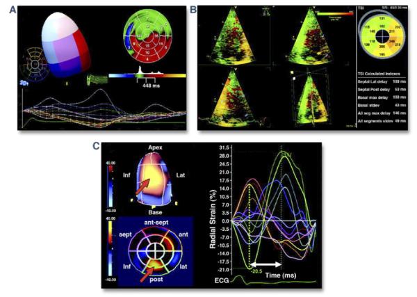
Figure 5. Three-Dimensional Echocardiographic Evaluation of Dyssynchrony.

(A) Systolic dyssynchrony index used to evaluate left ventricular (LV) mechanical dyssynchrony calculated from the standard deviation (stdev) of time to minimal systolic regional volume of the 17-segment LV model. (B) Time dispersion map relating relative regional delay to contraction of the left ventricle. The green regions represent earliest myocardial contraction, while orange and red regions are the latest to contract. (C) Three-dimensional speckle tracking for assessment of strain. The time dispersion to peak strain may be calculated (radial strain is shown). A polar map denoting relative regional delay (blue earliest, yellow latest) also provides visual assessment of dyssynchrony. ant = anterior; ECG = electrocardiogram; inf = inferior; lat = lateral; max = maximal; post = posterior; seg = segments; sept = septal; TSI = tissue synchronization index. Reprinted, with permission, from Delgado V, Bax JJ. Assessment of systolic dyssynchrony for cardiac resynchronization therapy is clinically useful. Circulation 2011;123:640-55.
