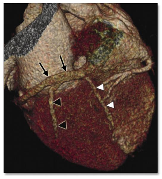
Figure 9. Volume-Rendered Image of the Coronary Sinus.

Volume-rendered cardiac computed tomographic image of the coronary sinus (black arrows). The posterior vein of the left ventricle (black arrowheads) and posterior interventricular vein (white arrowheads) may also be visualized. Reprinted, with permission, from Gopalan D, Raj V, Hoey ETD. Cardiac CT: noncoronary applications. Postgrad Med J 2010;86:165-73.
