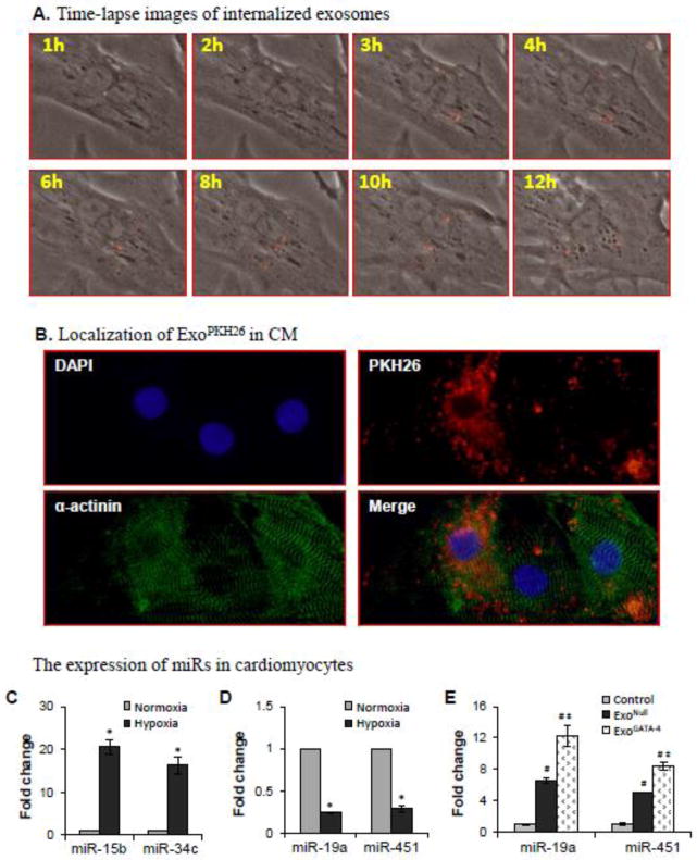
Figure 6.
The internalization of exosomes and the effect of internalized exosomes on the expression of miRs in CM. A, Time-lapse images following the addition of ExoPKH-26 to CM culture. B, Immunostaining shows the red fluorescence (ExoPKH26) was inside α-actinin-positive cells. C-E, Expression of miRs in CM after exposure to hypoxia for 24 hours and treatment with exosomes. *, p<0.05 vs normal control; #, p<0.05 vs hypoxia, and ‡, p<0.05 vs ExoNull treated, respectively.

