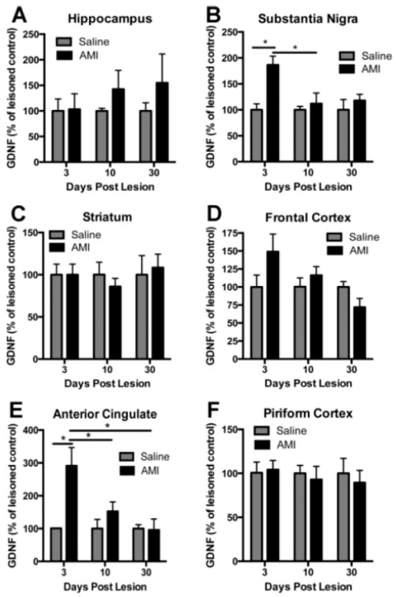
Figure 6. GDNF response to amitriptyline (AMI) post-lesion.

GDNF is differentially regulated in response to chronic antidepressant treatment and/or lesion. The region of interest was dissected and each hemisphere was analyzed independently with ELISA for GDNF. Adult rats were treated with saline or AMI (5mg/kg, i.p.) for 2 weeks prior to receiving 6-OHDA lesions on day 14. Subsets of these rats were sacrificed at 3, 10 or 30 days post-lesion. Levels of GDNF are depicted for each of the following regions: Hippocampus (A), substantia nigra (B), striatum (C), frontal cortex (D), anterior cingulate (E) and piriform cortex (F). Values are expressed as a percentage of saline lesioned controls. Data represent the mean+/-SEM and are combined from two separate experiments. *p<0.05, **p<0.001
