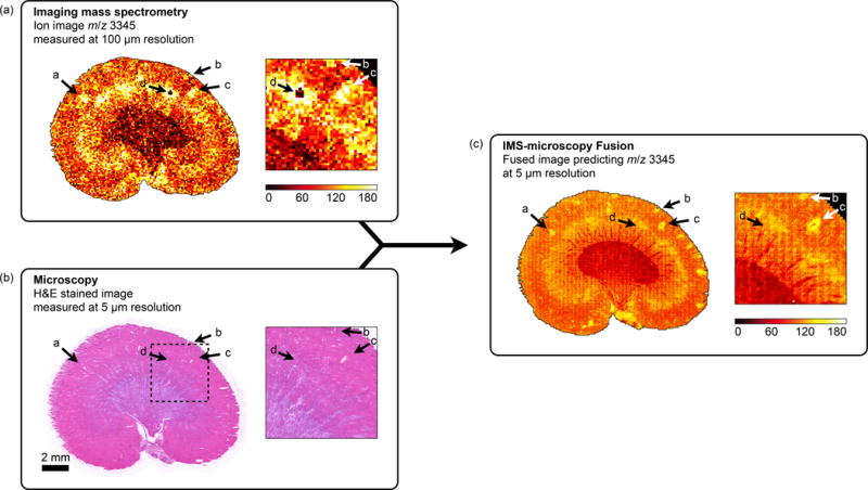
Figure 6.

Discovery of tissue features through multi-modal enrichment. An ion image for m/z 3,345 measured by IMS at 100 μm resolution in a rat kidney section (a) is fused with H&E stained microscopy acquired at 5 μm resolution (b) to produce an ion distribution prediction at 5 μm resolution (reconstr. score 85%) (c). Annotations a–c demonstrate multi-modal enrichment. If only IMS is considered, these features could be mistaken for ‘matrix hotspot’ noise. However, their successful propagation through the fusion process and their presence in the final fused image confirms they are genuine tissue features that are corroborated by another technology (in this case microscopy). If only microscopy is considered, these features are so faint that they would probably not be detected. Instead, they are only discovered by fusion with another data source. Annotation d demonstrates multi-modal attenuation. The lack of cross-modal support for this localized drop in ion intensity reduces confidence in the biological nature of this feature.
