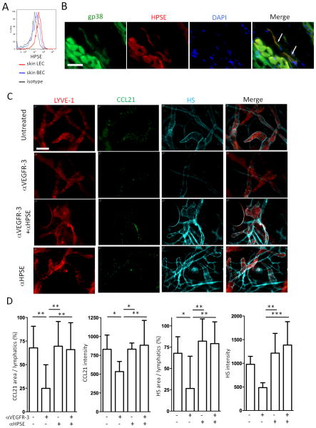
Figure 4. VEGFR-3 regulates HPSE.
(A) Ear pinnae single cell suspensions stained for CD45, CD31, gp38, and heparanase (HPSE) and analyzed by flow cytometry. Gating strategy is shown in Fig S1A. HPSE in LEC and BEC are shown. (B) Pinnae stained for HPSE, gp38, and DAPI. Original magnification 600×. Bar, 20μm. White arrows indicate LEC. (C) Control or anti-VEGFR-3 mAb (7μg) injected into pinnae along with anti-HPSE antibody (1μg) intradermally; ears harvested after 20 hours; and stained for LYVE-1 (Cy3), CCL21 (Dye Light 488), and heparan sulfate (HS, Alexa Fluor 647). Original magnification 200×. Bar, 100μm. (D) Same as (C), staining intensity of CCL21 and HS, and %CCL21- and %HS-positive area in lymphatics calculated. n = 10-12 fields/group. Results from 2 mice/group, 3 independent experiments. * p<0.05, ** p<0.005, *** p<0.0005 by one-way ANOVA.

