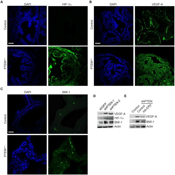
Figure 3. Role of ERβ repression in prostate tumorigenesis.
Ventral prostates of wild-type (control) and aged-matched Pten pc−/−mice were stained for (A) HIF-1α, (B) VEGF-A and (C) BMI-1 and analyzed by immunofluorescence microscopy. Scale bar, 50 µm. (D) Immunoblot showing the expression of HIF1α, VEGF-A and BMI-1 in PTEN-depleted (shPTEN) and control (shGFP) PNT1a cells. (E) Expression of VEGF-A and BMI-1 in ERβ expressing PTEN depleted cells (shPTEN-2+HA-ERβ) compared to PTEN depleted cells (shPTEN).

