Figure 5.
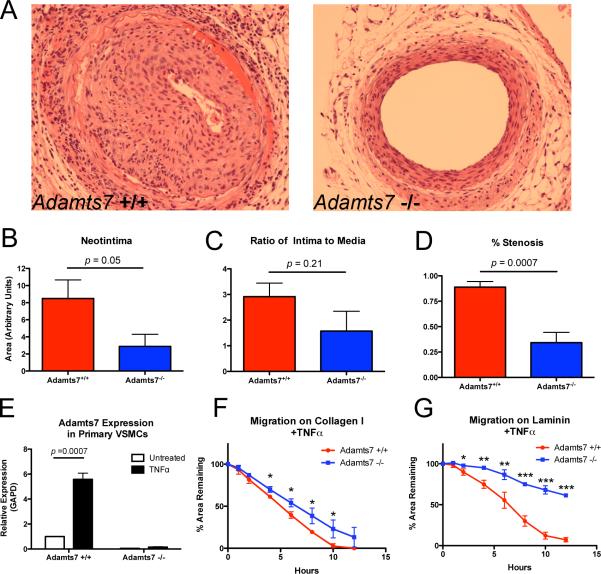
Neointima formation following femoral artery wire injury is attenuated by ablation of mouse Adamts7. (A) Wire injury was performed on femoral arteries of Adamts7−/−and WT animals (N=5 and 4, respectively), vessels were harvested at 28 days and neointima was assessed by H/E staining. Representative sections from WT and KO femoral arteries are shown. There was reduced (B) neointima (64%), (C) intima-to-media ratio (47%) and (D) percent stenosis (61%) in Adamts7−/− compared to WT. (E) Adamts7 expression measured by TaqMan real-time qPCR in primary VSMCs from Adamts7 WT or KO mice after treatment with 25ng/ml TNFα for 24-hours (N=3). (F,G) Migration of TNFα-stimulated Adamts7 KO primary VSMCs on collagen or laminin coated plates (N=3) is reduced compared to WT cells.

