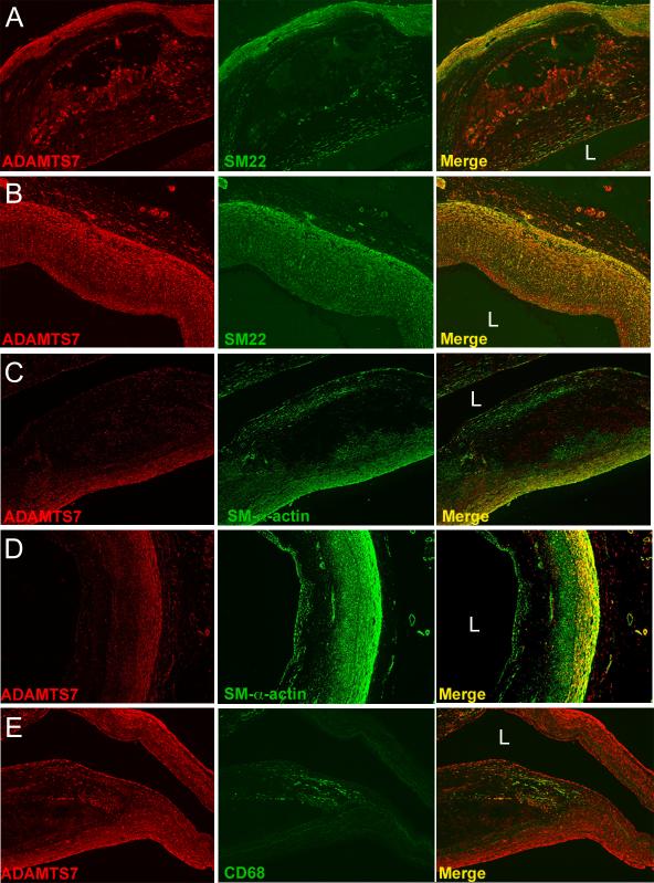
Figure 7.
ADAMTS7 colocalizes with VSMC markers in human atherosclerotic coronary artery sections. (A,B) Staining of human coronary artery lesions with antibodies directed towards ADAMTS7 and SM22, with merge of the two stains shown in the far right panels. (C, D) Staining of similar lesions shown in A and B for ADAMTS7 and SM-α-actin, with merge shown in far right columns. (E) Staining of the shoulder region of a human coronary artery lesion with antibodies to ADAMTS7 and CD68, with merge to the far right showing little overlap between the two stains. “L” denotes location of the lumen. Staining was performed in 12 different diseased human coronary artery samples, and representative images are shown.

