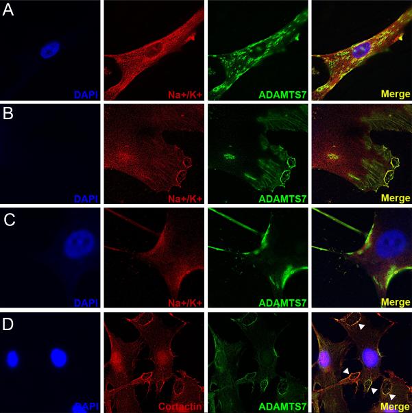
Figure 8.
ADAMTS7 localizes to the leading edge and podosome-like structures in primary human aortic smooth muscle cells (hAoSMCs). (A-C) immunofluorescent staining for ADAMTS7 and the cell surface marker Na+/K+ Ion Channel in primary human aortic SMCs. Each row is imaging of a different cell in the same experiment. (D) Co-staining for ADAMTS7 and cortactin, a marker of podosomes, in primary hAoSMCs. Arrowheads identify regions of strong overlap in focal adhesion-like structures.

